Configuration-specific Monoclonal Antibody Based
Arl3 Activation Assay Kit
Catalog Number:83001
20 assays
Product Description
Arl3 (Arf-like 3) is an ADP-ribosylation factors (Arf) family protein that differs from most Arf family members in the N-terminal extension. Nucleotide exchange of Arl3 is rapid and independent of lipids and detergents. Upon binding of GDT/GTP, Arl3 interacts with and regulates activities of series effector proteins, such as human retinal gene 4 (HRG4), δ-subunit of the cGMP phosphodiesterase (PDEδ), and binder of Arl2 (BART). Arl3 also binds microtubules in a regulated manner to alter specific aspects of cytokinesis via interactions with retinitis pigmentosa 2 (RP2). It has been proposed that RP2 functions in concert with Arl3 to link the cell membrane and the cytoskeleton in photoreceptors as part of the cell
signaling or vesicular transport machinery.
Currently there is no direct assay to measure the activation of Arl3 GTPases.
Bioyears Biosciences Arl3 Activation Assay Kit is based on the configuration-specific monoclonal
antibody that specifically recognizes Arl3-GTP, but not Arl3-GDP. Given the high affinity of
monoclonal antibodies to their antigens, the activation assay could be performed in a short time. This assay provides the reliable results with consistent reproducibility.
These anti Arl3 GTP monoclonal antibodies can also be used to monitor the activation of Arl3 in cells
and in tissues by immunohistochemistry.
Bioyears Biosciences Arl3 Activation Assay Kit provides a simple and fast method to monitor the activation of Arl3. Each kit provides sufficient quantities to perform 20 assays.
Assay Principle
Bioyears Biosciences Arl3 Activation Assay Kit bases on the configuration-specific anti Arl3-GTP monoclonal antibody to measure the active Arl3-GTP levels, either from cell extracts or from in vitro GTPγS loading Arl3 activation assays. Briefly, anti active Arl3 mouse monoclonal antibody will be incubated with cell lysates containing Arl3-GTP. The bound active Arl3 will then be pulled down by protein A/G agarose. The precipitated active Arl3 will be detected by immunoblot analysis using anti Arl3 rabbit polyclonal antibody.
Kit Components
1. Anti active Arl3, Mouse Monoclonal Antibody (Catalog No. 26925): One vial – 22 µL (1
mg/ml) in PBS, pH 7.4, containing 50% glycerol and 0.05% sodium azide. This antibody
specifically recognizes Arl3-GTP from all vertebrates.
2. Protein A/G Agarose (Catalog No. 30301): One vial – 400 µL of 50% slurry.
3. 5X Assay/Lysis Buffer (Catalog No. 30302): One bottle – 30 mL of 250 mM Tris-HCl, pH 8, 750mM NaCl, 50 mM MgCl2, 5 mM EDTA, 5% Triton X-100.
4. Anti Arl3, Mouse monoclonal Antibody (Catalog No. 070): 26 One vial – 100 µL (1mg/ml)
in PBS, pH 7.4, contained 50% glycerol.
5. 100 X GTPγS (Catalog No. 30303): One vial –100 µl at 10 mM, use 5 µL of GTPγS for
GTP-labeling of 0.5 mL of cell lysate.
6. 100 X GDP (Catalog No. 30304): One vial –100 µl at 100 mM, use 5 µL of GDP for
GDP-labeling of 0.5 mL of cell lysate.
Storage
Store all kit components at 4ºC until their expiration dates.
Materials Needed but Not Supplied
1. Stimulated and non-stimulated cell lysates
2. Protease inhibitors
3. 4 °C tube rocker or shaker
4. 0.5 M EDTA, pH8.0
5. 1 M MgCl2
6. 2X reducing SDS-PAGE sample buffer
7. Electrophoresis and immunoblotting systems
8. Immunoblotting wash buffer such as TBST (10 mM Tris-HCl, pH 7.4, 0.15 M NaCl, 0.05%
Tween-20)
9. Immunoblotting blocking buffer (TBST containing 5% Non-fat Dry Milk or 3% BSA)
10. PVDF or nitrocellulose membrane
11. Secondary Antibody
12. ECL Detection Reagents
Reagent Preparation
• 1X Assay/Lysis Buffer: Mix the 5X Stock briefly and dilute to 1X in deionized water. Just prior to usage, add protease inhibitors such as 1 mM PMSF, 10 µg/mL leupeptin, and 10 µg/mL aprotinin.
Sample Preparation
Adherent Cells
1. Culture cells (one 10-cm plate, ~ 107cells) to approximately 80-90% confluence. timulate cellswith activator or inhibitor as desired.
2. Aspirate the culture media and wash twice with ice-cold PBS.
3. Completely remove the final PBS wash and add ice-cold 1X Assay/Lysis Buffer to the cells (0.5- 1 mL per 10 cm tissue culture plate).
4. Place the culture plates on ice for 10-20 minutes.
5. Detach the cells from the plates by scraping with a cell scraper.
6. Transfer the lysates to appropriate size tubes and place on ice.
7. If nuclear lysis occurs, the cell lysates may become very viscous and difficult to pipette. If this occurs, lysates can be passed through a 27½-gauge syringe needle 3-4 times to shear the genomic DNA.
8. Clear the lysates by centrifugation for 10 minutes (12,000 x g at 4 °C).
9. Collect the supernatant and store samples (~1-2 mg of total proteins) on ice for immediate use, or snap freeze and store at - 70 °C for future use.
Note: In vivo stimulation of cells will activate approximately 10% of the available Arl3, whereas in vitro GTPγS protein loading will activate nearly 90% of the Arl3.
1. Aliquot 0.5 ml of each cell extract to two microfuge tubes (or use 1 µg of purified Arl3 protein).
2. To each tube, add 20 µl of 0.5 M EDTA (to 20 mM final concentration).
3. Add 5 µl of 100 X GTPγS (to 100 µM, final concentration) to one tube (positive control).
4. Add 5 µl of 100 X GDP (to 1 mM, final concentration) to the second tube (negative control).
5. Incubate the tubes at 30°C for 30 minutes with agitation.
6. Stop loading by placing the tubes on ice and adding 32.5 µl of 1 M MgCl2 (to 60 mM, final concentration).
Assay Procedure
I. Active Arl3 Pull-Down Assay
1. Aliquot 0.5 – 1 mL of cell lysate to a microcentrifuge tube.
2. Adjust the volume of each sample to 1 mL with 1X Assay/Lysis Buffer.
3. Add 1 µl anti active Arl3 monoclonal antibody to the tube.
4. Thoroughly resuspend the protein A/G Agarose bead slurry by vortexing or titurating.
5. Quickly add 20 µL of resuspended bead slurry to each tube.
6. Incubate the tubes at 4 °C for 1 hour with gentle agitation.
7. Pellet the beads by centrifugation for 1 min at 5,000 x g.
8. Aspirate and discard the supernatant, making sure not to disturb/remove the bead pellet.
9. Wash the bead 3 times with 0.5 mL of 1X Assay/Lysis Buffer, centrifuging and aspirating each
time.
10. After the last wash, pellet the beads and carefully remove all the supernatant.
11. Resuspend the bead pellet in 20 µL of 2X reducing SDS-PAGE sample buffer.
12. Boil each sample for 5 minutes.
13. Centrifuge each sample for 10 seconds at 5,000 x g.
II. Electrophoresis and Transfer
1. Load 15 µL/well of pull-down supernatant to a polyacrylamide gel (17%). Also, it’s
recommended to include a pre-stained MW standard (as an indicator of a successful transfer in step 3).2. Perform SDS-PAGE following the manufacturer’s instructions.
3. Transfer the gel proteins to a PVDF or nitrocellulose membrane following the manufacturer’s instructions.
III. Immunoblotting and Detection (all steps are at room temperature, with agitation)
1. Following the electroblotting step, immerse the PVDF membrane in 100% Methanol for 15 seconds, and then allow it to dry at room temperature for 5 minutes.
Note: If Nitrocellulose is used instead of PVDF, this step should be skipped.
2. Block the membrane with 5% non-fat dry milk or 3% BSA in TBST for 1 hr at room temperature with constant agitation.Incubate the membrane with anti Arl3 polyclonal
antibody, freshly diluted 1:50~1000
(depending on the amount of Arl3 proteins in your samples) in 5% non-fat dry milk or 3%BSA/TBST, for 1-2 hr at room temperature with constant agitation or at 4
oC overnight.
3. Wash the blotted membrane three times with TBST, 5 minutes each time.
4. Incubate the membrane with a secondary antibody (e.g. Goat Anti Rabbit IgG, HRP- conjugate),freshly diluted 1:1000 in 5% non-fat dry milk or 3% BSA/TBST, for 1 hr at room temperaturewith constant agitation.
5. Wash the blotted membrane three times with TBST, 5 minutes each time.
6. Use the detection method of your choice such as ECL.
Example of Results
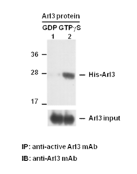
The following figure demonstrates typical results seen with Bioyears Biosciences Arl3 Activation Assay Kit. One should use the data below for reference only.

Arl3 activation assay. Purified His-tagged Arl3 proteins (Cat. #10152) were mmunoprecipitated with the anti active Arl3 monoclonal antibody (Cat. #26925) after treated with GDP (lane 1) or GTPγS (lane 2), and was blotted with anti Arl3 monoclonal antibody(Cat. #070). 26 Input control is shown in bottom panel.


 电话: 027-87561633
 邮箱: Bioyears@126.com
 地址: 武汉市东湖开发区关东科技工业园3#产业区3-3栋15号